
Doctors use nuclear stress testing to see blood flow, and the test highlights areas with poor circulation. A small amount of radioactive tracer helps create the images the cardiologist needs to review. Because the substance travels through your arteries, a special camera detects the energy it releases as it passes nearby. Here is more information about the role of nuclear stress testing in heart disease diagnosis:
Evaluating Cardiovascular Health
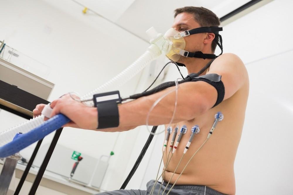
If you experience chest pain or shortness of breath, this procedure provides details about blood flow and heart health. The test identifies blockages in arteries, and it assesses damage from previous attacks on the heart muscle. Your physician can order this exam to check for coronary artery disease or other related cardiovascular issues. You might walk on a treadmill, or a provider will administer medication to simulate exercise stress.
The goal is to measure the heart’s ability to handle stress under controlled clinical conditions. When standard exercise tests do not provide enough data, this method offers more insight into your health. The results guide future treatment plans, or they confirm the effectiveness of the current medications you take daily. It determines if your heart receives enough blood during physical activity or periods of higher stress.
Showing Blood Flow
The tracer mixes with your blood, and the camera tracks its path through vessels in the body. Cold spots indicate areas where blood supply is inadequate or completely blocked due to arterial narrowing. As the material moves through your system, it accumulates in areas that have good blood flow retention. The images display distribution patterns clearly, so the doctor sees exactly where the blood travels.
Standard uses include:
- Identifying blocked coronary arteries.
- Assessing heart valve function.
- Measuring heart chamber size.
Creating Detailed Images
The camera rotates around your chest to capture images from multiple angles for a complete view. These slices form a 3D model of the heart, and doctors view them on a high-resolution monitor. Because the technology is advanced, it creates slices of images for review by a trained cardiologist. This technology allows for a non-invasive look inside the structure of your cardiovascular system.
While the scan takes time, the detail provided surpasses standard X-rays significantly in precision. Providers compare images taken during rest with those taken during stress to find specific changes. A difference between the two sets appears on the screen, or the images look exactly the same. If the images match perfectly, it suggests that blood flow remains steady during physical exertion.
Healthy tissue absorbs the tracer easily, but damaged tissue absorbs very little of the radioactive substance. When scar tissue is present, it does not conduct electricity or pump blood well through the heart. This contrast allows for the precise identification of scars left behind by previous events. The cardiologist reviews these specific areas, and they determine the extent of the permanent damage.
Diagnosing Heart Conditions
Accurate diagnosis guides the management of complex cardiac issues found during the nuclear stress testing process. Some patients require surgery immediately, but others manage their condition with lifestyle changes and medication alone. If the test reveals significant blockages, your doctor may recommend additional treatment. This helps prevent future heart attacks by addressing the specific problem areas found during the exam.
Schedule Nuclear Stress Testing
Once you understand the procedure, you can be more prepared for your upcoming appointment at our clinic. Early detection aids treatment options, so do not delay your cardiovascular care for another day. Contact a cardiology clinic today to book your nuclear stress test with our experienced medical staff.
Leave a Reply