
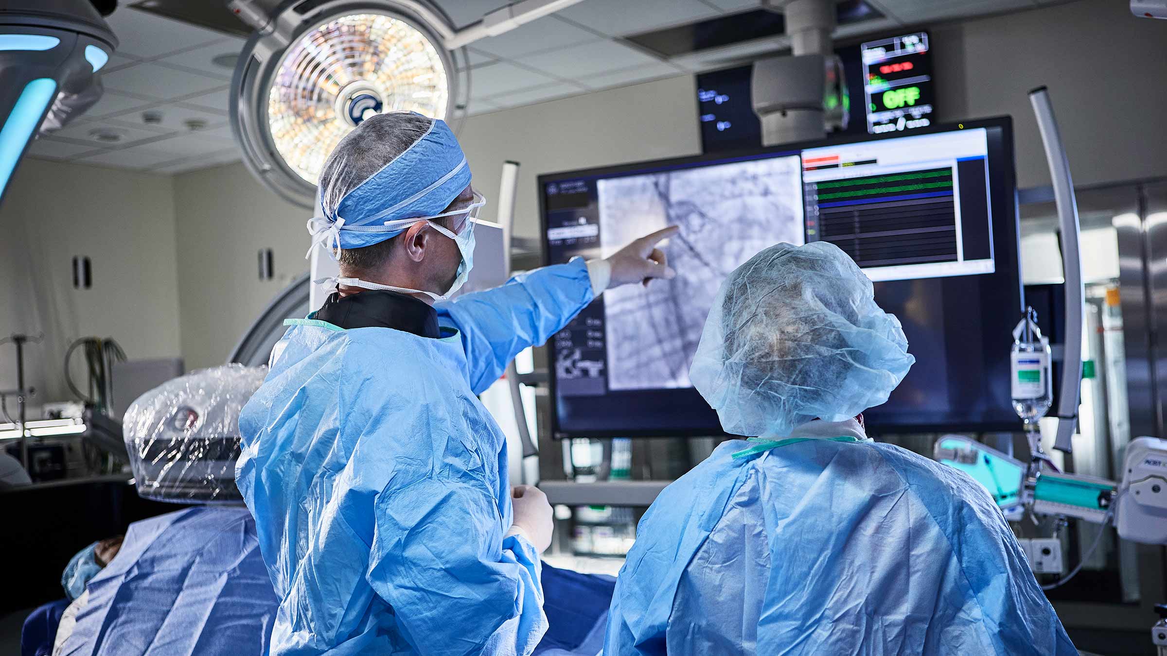
While traditional surgery is one option for vascular conditions, another method involves minimally invasive procedures. This field is known as interventional radiology. Interventional radiologists use medical imaging, like X-rays and CT scans, to guide small instruments through the body. Here is how this approach allows them to diagnose and treat conditions without large incisions:
Embolization
Embolization is a procedure that blocks blood flow to a specific area of the body. Doctors perform this treatment to stop bleeding or cut off the blood supply to abnormal growths, such as uterine fibroids or certain tumors. Using imaging for guidance, an interventional radiologist typically inserts a thin tube called a catheter into a blood vessel, often through a tiny opening in the groin or wrist.
The radiologist carefully guides the catheter to the target location. Once it reaches the destination, the radiologist injects a substance through the catheter. This substance creates a blockage in the vessel. The choice of material depends on the specific condition and whether the blockage needs to be temporary or permanent. This targeted approach affects only the intended blood vessels and leaves the surrounding healthy tissue untouched.
Radiofrequency Ablation
Radiofrequency ablation (RFA) is another minimally invasive interventional radiology treatment that uses heat generated by an electrical current to destroy abnormal cells. Doctors often use this technique to treat varicose veins and certain types of tumors. The interventional radiologist begins the process by numbing the skin over the treatment area.
Using ultrasound imaging, the doctor guides a needle-like probe through the skin and into the targeted tissue. When the probe is correctly positioned, it sends out a high-frequency electrical current that heats and destroys the problematic cells. For varicose veins, the heat causes the vein to close, and blood naturally reroutes through healthier veins. Over time, the body absorbs the treated vein.
Angioplasty and Stents
When arteries become narrow or blocked due to plaque buildup, blood flow becomes restricted. This condition can cause severe health issues. Doctors use angioplasty to open these blocked arteries and restore proper blood circulation.
During an angioplasty, an interventional radiologist inserts a catheter with a small balloon at its tip into an artery. They guide the catheter to the blockage using imaging. Then, they inflate the balloon, pressing the plaque against the artery wall and widening the vessel.
In many cases, doctors place a stent during the angioplasty. A stent is a small, mesh-like tube that helps keep the artery open after removing the balloon. It acts as a scaffold to support the artery wall. Some stents contain medication that slowly releases to prevent the artery from narrowing again.
Schedule Interventional Radiology Treatments
Understanding the alternatives to surgery is a positive first step. Interventional radiology offers minimally invasive procedures that address a range of vascular issues with precision. If you believe you might benefit from one of these treatments, discussing your situation with a specialist can provide clarity. A radiologist can help you navigate your options and understand what might be right for your health needs. Contact a clinic today to schedule a consultation and learn more about interventional radiology services.
Leave a Reply